
Medical imaging AI becomes far more useful when it is integrated directly into the tools that clinicians, researchers, and imaging teams already use.
That is the direction we are exploring with DICOM Vision®: bringing AI-assisted workflows into a DICOM-native environment, reducing the need to move data between disconnected PACS systems, scripts, viewers, conversion tools, and annotation platforms.
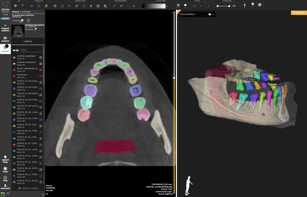
In this short technical demo, we show an early integration of AI-powered dental segmentation inside DICOM Vision®, using TotalSegmentator and ToothFairy to automatically segment teeth and dental structures from CT/DICOM imaging data.
The demo is intentionally short, around one minute, but it highlights an important concept: AI segmentation should not be an isolated post-processing step. It should become part of the imaging workflow itself.
From remote PACS to AI segmentation
Traditional segmentation workflows often require several manual steps:
1 – Export the DICOM study from a PACS or viewer.
2 – Anonymize or prepare the data manually.
3 – Convert or preprocess the imaging files.
4 – Run an external AI model or script.
5 – Export the segmentation result.
6 – Re-import the output into another viewer or annotation tool.
7 – Manually inspect, refine, or use the result for downstream work.
This process can work for research, but it creates friction. Every additional tool, file conversion, manual export, and data-handling step increases complexity, slows down iteration, and introduces potential privacy or compliance risks.
With DICOM Vision®, the workflow can be much cleaner.
Imaging data can be imported directly from a remote PACS, with anonymization applied during the process. From there, the study remains inside a DICOM-oriented environment, while AI segmentation can be launched, visualized, and reviewed directly from the platform.
The workflow becomes:
Remote PACS → anonymized import → DICOM Vision® → AI segmentation → visualization → review → annotation/dataset preparation
This creates a more controlled path for teams working on medical imaging research, AI model development, data preparation, or advanced visualization.
Why dental segmentation matters
Dental and maxillofacial imaging often involves complex anatomical structures, high-resolution CT or CBCT data, and time-consuming manual segmentation tasks.
Automatic teeth segmentation can support several non-diagnostic workflows, including:
- anatomical visualization;
- annotation assistance;
- dataset preparation;
- AI training data generation;
- research workflows;
- pre-processing for downstream models;
- quality control of segmentation pipelines;
- comparison between manual and AI-assisted segmentation.
Manual segmentation of dental structures can be slow and repetitive. AI-assisted segmentation does not remove the need for expert review, but it can provide a useful starting point and significantly reduce the amount of manual work required.
TotalSegmentator and ToothFairy
This demo combines DICOM Vision® with external AI segmentation models, including TotalSegmentator and ToothFairy-related dental segmentation capabilities.
The purpose of the integration is not to replace expert review. The purpose is to make AI output easier to access, visualize, inspect, and use inside a structured imaging workflow.
For a medical imaging platform, model integration is only one part of the problem. The other part is workflow integration:
- how the input study is selected;
- how the model is executed;
- how the segmentation result is stored;
- how the result is visualized;
- how users inspect the output;
- how annotations are exported or reused;
- how the workflow fits into existing DICOM/PACS environments.
This is where DICOM Vision® focuses: making AI tools usable within practical imaging workflows.
Integrated AI workflows instead of disconnected tools
One of the main challenges in medical AI adoption is not only model performance. It is usable.
A model can be technically impressive, but if using it requires manual exports, command-line scripts, format conversions, and isolated viewers, adoption becomes harder.
Integrated workflows can help reduce this gap.
With DICOM Vision®, we aim to connect:
- remote PACS import;
- DICOM anonymization;
- DICOM viewing;
- PACS-oriented workflows;
- AI-assisted segmentation;
- annotation tools;
- collaborative review;
- dataset creation;
- validazione e iterazione di modelli AI.
For research and AI development teams, this means that data acquisition, anonymization, segmentation, review, and dataset preparation can become part of the same controlled workflow, instead of being split across multiple tools and manual steps.
Early demo, not a diagnostic tool
This video is an early technical demonstration. It is focused on workflow validation, technical integration, and visualization of AI-generated segmentation results. It is not intended to demonstrate diagnostic performance, clinical decision-making, or autonomous medical interpretation.
Any AI-generated segmentation output should be reviewed by qualified users before being used in research, validation, or clinical contexts.
What this enables
Even at this early stage, this type of integration opens useful possibilities:
- faster segmentation workflows;
- reduced manual preparation time;
- easier dataset creation;
- better visualization of anatomical structures;
- smoother AI model evaluation;
- improved collaboration between imaging, AI, and clinical teams;
- easier comparison between automated and manually refined annotations.
For teams building or validating medical AI systems, these workflow improvements can be as important as the model itself.
The direction for DICOM Vision®
DICOM Vision® is evolving toward a platform where medical imaging, AI assistance, annotation, and collaboration are part of the same workflow.
This dental segmentation demo is one example of that direction.
We are continuing to explore integrations that make AI more usable in real medical imaging environments, especially for:
- segmentation;
- study analysis;
- triage;
- dataset preparation;
- annotation
- AI validation;
- DICOM/PACS interoperability.
AI in medical imaging should not live only in notebooks, isolated scripts, or disconnected research tools.
It should be accessible where imaging work already happens.
That is what we are building with DICOM Vision®.